Comparing AFB and LED-Fluorescence Microscopy
ContentThe comparison of the Acid-Fast Bacilli (AFB) and Light Emission Diode (LED) Fluorescence Microscopy is tabulated below:
Direct Microscopy for Acid Fast Bacilli (AFB)
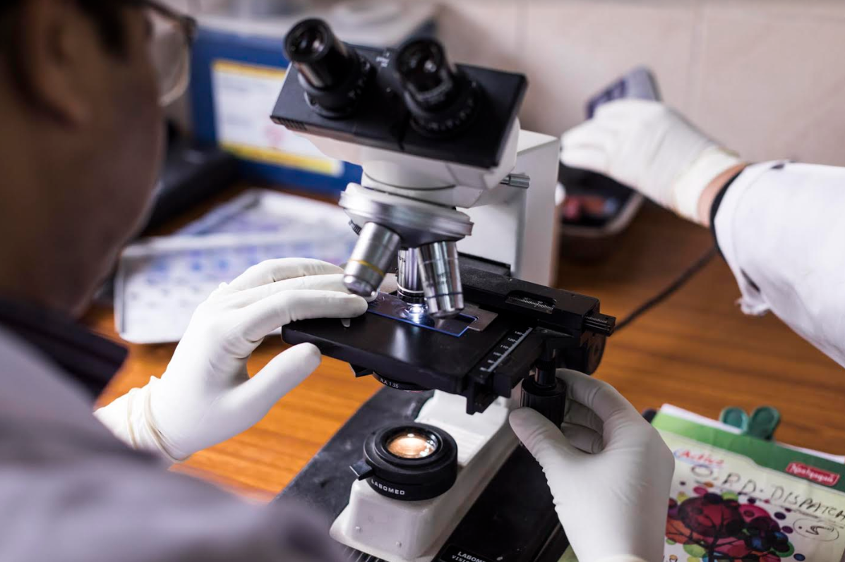
Figure 1: Bright Field Microscope used in Ziehl–Neelsen (ZN) staining of AFB
Light Emission Diode (LED)
Fluorescence Microscopy
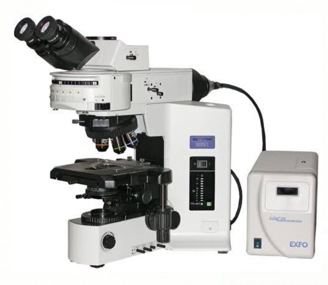
Figure 2: LED-Fluorescent Microscope used in fluorescent staining of AFB
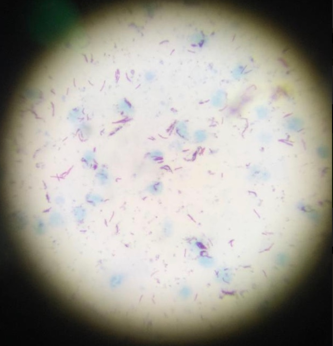
Figure 3: ZN-stained slide used in direct microscopy

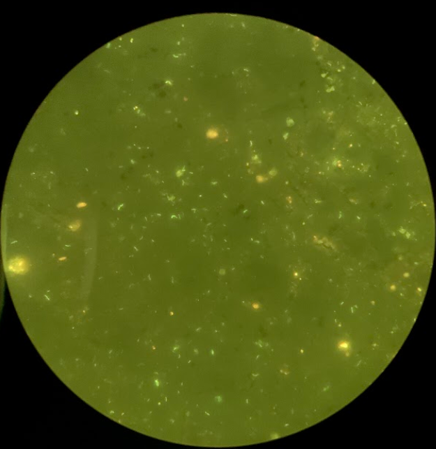
Figure 4: Auramine-O-stained slide used in fluorescent microscopy
Method used widely for the diagnosis and confirmation of pulmonary tuberculosis
Method used less commonly for the diagnosis and confirmation of pulmonary tuberculosis
Staining procedure complex
Staining procedure simpler
Examination at lower magnification is NOT possible
Examination at lower magnification is possible
Takes more time than LED-FM
Takes 75% less time than ZN and chances to lose scanty slides are also minimized.
Low durability
High durability
Less sensitive
10% more sensitivity than Bright Field ZN microscopy
As specific as LED-FM
Equally as specific as ZN microscopy
No need for an experienced lab technician
Needs an experienced lab technician
Technical errors are common
Technical errors are less common
Misses the paucibacillary TB cases, especially when the patient is co-infected with HIV
Identifies the paucibacillary TB cases especially when the patient is co-infected with HIV
Resources
- WHO Policy Statement - Fluorescence Light Emitted Microscope for the Diagnosis of TB, 2010.
- A Comparative Study of Auramine Staining using LED Fluorescent Microscopy with Ziehl- Neelsen Staining in the Diagnosis of Pulmonary Tuberculosis, Journal of Evolution of Medical and Dental Sciences, Volume 2, Issue 20, May 2013.
- Comparison of Direct versus Concentrated Smear Microscopy in Detection of Pulmonary Tuberculosis, BMC Res Notes, 2013; 6 : 291.
Kindly provide your valuable feedback on the page to the link provided HERE
Ideal Infrastructure Required for Microscopy Centers
ContentUnder the National Tuberculosis Elimination Program (NTEP) network, the most peripheral laboratories are the Designated Microscopy Centres (DMC) which serve a population of around 100,000 each (50,000 population norm in tribal and hilly areas).
The basic requirements for microscopy centres include (Figure):
- Good ventilation by exhaust fan and open windows
- A table/ work bench to prepare smears
- A sink to stain smears
- A table/ bench to examine smears
- A table/ bench for documentation
- Basin for hand washing with supply of water
- Good lighting
- Non-slip flooring
- Sample receiving area
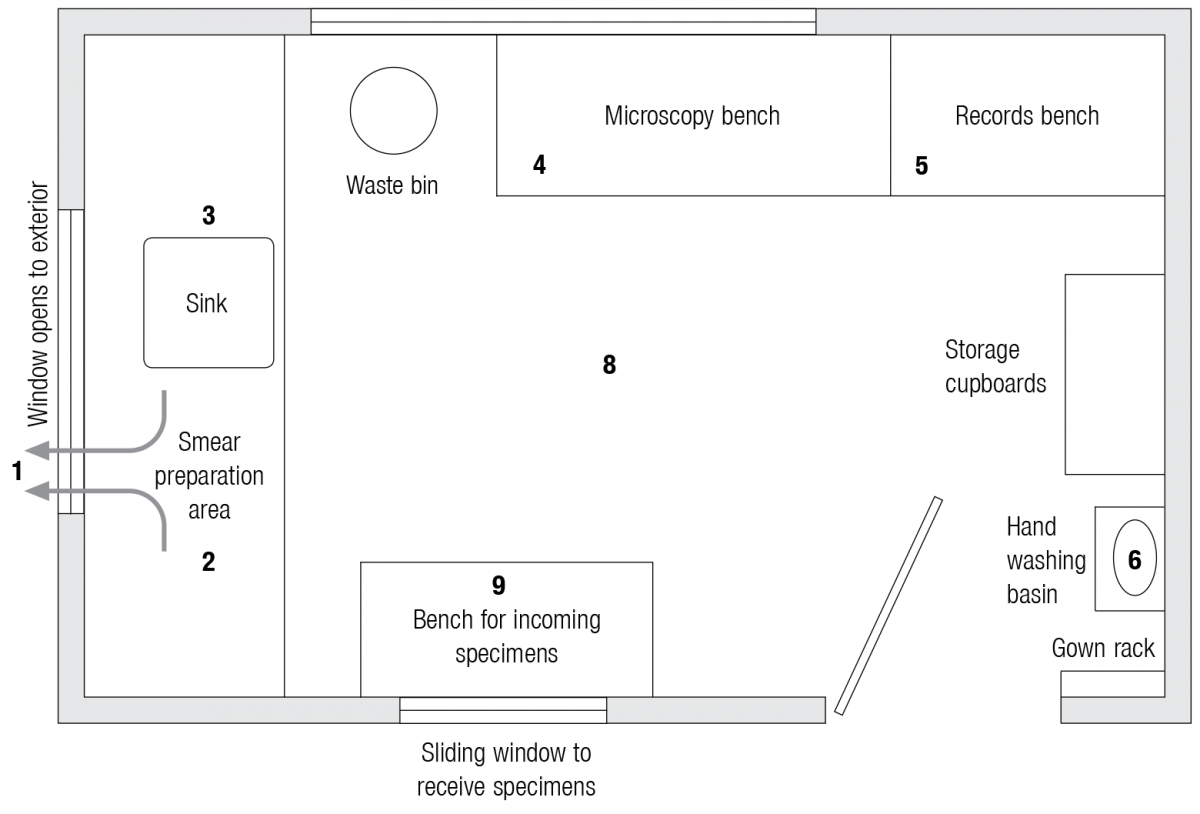
Figure: Ideal Infrastructure required for Microscopy Centres
Laboratory Diagnosis of Tuberculosis by Sputum Microscopy
ESTABLISHMENT
PROVISIONS UNDER NTEP FOR DMC FACILITIES
Infrastructure development
Normative amount as per “Norms and basis of costing” for NTEP. Unit cost for initial establishment/ refurbishment/ upgradation/ maintenance of civil work to be carried out as per rates prescribed by Public Works Department (PWD) or cell/ division/ corporation/ wing for infrastructure development
Human resources
NTEP trained Lab Technician (LT)
Diagnosis
- Provision for sputum collection in the presence or absence of an LT
- Adequate space for storing and using binocular microscope, essential lab consumables, space for recording and reporting
- Availability of running water
- Availability of electricity
- Availability of biomedical waste management facilities
Equipment
Binocular microscope with a light source
Location of DMC
Ideally, the DMC should be located in a peripheral health facility that has at least 50 new adult OPD/day.
Resources
- Technical and Operational Guidelines for TB Control in India, 2016.
- Laboratory Diagnosis of Tuberculosis by Sputum Microscopy, GLI Initiative.
Kindly provide your valuable feedback on the page to the link provided HERE
Microscopy
ContentMicroscopy is a TB diagnostic technology that utilizes the acid-fastness property of Mycobacterium tuberculosis to visualize it under a microscope. Results of sputum smear microscopy can either be smear-negative, or smear-positive (with various grades).
Advantages:
- It is currently the most accessible and cheapest TB diagnostic test available under National TB Elimination Programme (NTEP) in India.
- It has the shortest turnaround time for diagnosis.
- It has high specificity.
Limitations:
- Low sensitivity. It becomes positive only when more than 5000 bacilli/ml of sample are present. Hence, cases would be missed in early disease, or when an inappropriate biological specimen is provided, where bacterial load in sputum is less.
- It is unable to differentiate between M. tuberculosis and Non-tuberculous Mycobacteria (NTM). This is predominantly an issue in geographies with lower burden.
There are two types of microscopies used in NTEP: Ziehl-Neelsen (ZN) Microscopy and Fluorescence Microscopy (FM). These vary in the type of stain and microscope used. FM is newer of the two types and is currently recommended for use over ZN.
Resources
Parts of a Microscope
ContentVideo fileWorking with a Microscope
ContentVideo file
Fullscreen